Sunday, October 14, 2007

Treatment of Anisometropic Amblyopia in Children with Refractive Correction.

Introduction

Amblyopia is a frequent cause of monocular vision loss in children. A difference in refractive error between the two eyes (anisometropia) is a common cause of amblyopia, being present as the only identifiable amblyogenic factor in 37% of cases and present concomitantly with strabismus in an additional 24% of clinical populations.

Although there have been reports that refractive correction alone results in improved vision in anisometropic amblyopia, it is generally held that the majority of cases will need additional treatment because refractive correction alone will not be sufficient to completely treat the amblyopia. Thus, patching or pharmacological treatment is often prescribed simultaneously or soon after the refractive correction is provided.

Discussion

In this prospective observational study of 84 untreated anisometropic amblyopic children 3 to less than 7.

Visual acuity improvement with refractive correction alone in children with anisometropic amblyopia has been observed in both retrospective and pilot studies. However, only recently has the magnitude and time course of this phenomenon been evaluated prospectively. Stewart and colleagues found a mean improvement of nearly 3 lines in 18 patients with anisometropic amblyopia treated with spectacles. They have termed this effect “refractive adaptation,” but we prefer to refer to it as optical treatment of amblyopia.

The time course in our study for improvement to best amblyopic eye visual acuity was variable. The majority (83%) of patients stopped improving before 15 weeks, but one patient improved for 30 weeks. In the study of Stewart et al., improvement occurred for an average of 15.6 weeks in their subgroup of patients with anisometropic amblyopia.

The finding of visual acuity improvement over many weeks for children with anisometropic amblyopia treated with refractive correction alone can guide (1) clinicians on the expected duration of treatment when using spectacles alone as the treatment and (2) investigators who want to control for the treatment effect of refractive correction when evaluating the effectiveness of other amblyopia treatments. In earlier randomized trials of patching and pharmacological treatment conducted by PEDIG, optimum spectacle correction was required for a minimum of 4 weeks prior to enrollment. The results of the present study suggest that in these previous trials, some of the visual acuity improvement was due to optical treatment of amblyopia with spectacles. Although this would have the effect of reducing the statistical power to detect a treatment group difference, these trials were highly powered and this effect of spectacles therefore should not alter the primary conclusions of those studies.

Patients who stopped improving based on serial visual acuity measurements were enrolled in the control group of the randomized trial (complete results reported in a companion paper). We were surprised that a substantial proportion of these patients (62%) demonstrated further improvement (1 line or greater) of amblyopic eye visual acuity after another 5 weeks of spectacle wear, and some continued to show further improvement 10 and 15 weeks from randomization. Therefore, the criterion that we used in this study to define visual acuity stabilization (i.e., less than 1 line improvement from the prior 5-week visit on at least two tests of acuity) was not sufficient.

There are several possible explanations for the continued improvement after apparent stability. These include the possibility of test-retest variability including poor test performance on that particular day and the inability to detect acuity improvements of less than one logMAR line with the ATS visual acuity protocol. It is also possible that visual acuity improvement may temporarily plateau for a time period and then subsequently improve again. Based on these considerations, one may want to wait for two or perhaps even three 5-week follow-up cycles with no further improvement before assuming that all acuity improvement has been obtained from the wearing of the spectacle lenses. Alternatively, the clinician might choose to lengthen the time interval between visits to 10 or 12 weeks.

We evaluated the influence of age, degree of anisometropia, and baseline amblyopic eye visual acuity on the response to treatment. The beneficial effect of spectacle correction was consistent throughout the 3 to less than 7 years age range. We are not aware of other studies that have evaluated the association of age at presentation with treatment outcome for patients with anisometropic amblyopia who were treated with spectacles alone. Regarding other patient characteristics, better baseline acuity in the amblyopic eye was related to an increased chance for resolution of amblyopia. Severe amblyopia was unlikely to resolve with spectacle correction alone. Higher rates of resolution were associated with lesser amounts of anisometropia. Again, we are not aware of other studies that have evaluated the relationship of baseline visual acuity or magnitude of anisometropia with treatment outcome for children with anisometropic amblyopia treated with spectacle correction alone.

In translating our results into clinical practice, the findings must be viewed in the context of the clinical profile of the cohort enrolled into the study: children 3 to less than 7.

Based on this study of untreated 3 to less than 7 year old children with anisometropic amblyopia, one can expect most patients treated with refractive correction alone to have two lines or more of improvement in visual acuity, while at least one third will actually experience resolution of amblyopia if treated long enough with spectacles. Although anisometropic amblyopic children with severe amblyopia and /or higher degrees of anisometropia are much less likely to resolve with optical correction alone, many such patients could begin their prescribed occlusion or pharmacological regimen with better vision if first treated with spectacles alone. This, in turn, might decrease the burden of treatment on the child and parents and enhance compliance with patching treatment, because denser levels of amblyopia have been reported to be associated with poorer treatment compliance.

Conclusion:

Refractive correction alone improves visual acuity in many cases and results in resolution of amblyopia in at least one third of 3 to less than 7-year-old children with untreated anisometropic amblyopia. While most cases of resolution occur with moderate (20/40 to 20/100) amblyopia, the average 3-line improvement in visual acuity resulting from treatment with spectacles may lessen the burden of subsequent amblyopia therapy for those with denser levels of amblyopia.

Saturday, October 13, 2007

Reading with optical magnifiers: page navigation strategies and difficulties.

Introduction

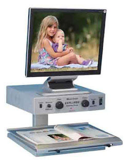
Optical magnifiers, such as hand and stand magnifiers are commonly prescribed to assist visually impaired people with reading. The task for which a magnifier is used may vary from spot reading, such as finding the total on a utility bill, to fluent reading of a newspaper or a novel. Due to the restricted field of view, the magnifier has to be moved to locate the required information (e.g., the total on the utility bill) or the start of a new line, and/or read a line of text. Good control and the adoption of efficient patterns of magnifier movement are fundamental to the successful use of both hand and stand magnifiers. Clinical experience suggests that some patients have difficulty with navigating a magnifier around the page, reporting that they miss lines or repeat lines when reading. An understanding of how visually impaired readers actually use their hand and stand magnifiers is relevant to the development of improved methods of training.

Previous studies of magnifier movements have either used fully-sighted participants reading with optical magnifiers or visually impaired participants reading with closed-circuit television (CCTV) systems; to our knowledge magnifier movements of visually impaired readers using optical magnifiers have not been investigated. Optical magnifiers tend to be prescribed more commonly than CCTVs and the method of using an optical magnifier is very different from that of a CCTV. In the study by Neve, hand magnifiers were moved across a glass plate, which was positioned parallel to and at a fixed distance from the text; thus the participants did not have to maintain the magnifier at the correct distance from the page, which removed one of the usual requirements of using a hand magnifier.

Discussion

Magnifier movement patterns

When reading along a line (forward phase) participants primarily used either a straight or diagonal downward movement. A straight movement, keeping the vertical positioning of the magnifier constant across the line, is the movement pattern that we expected for the forward phase; the downward movement was surprising. The vertical displacement per line in the forward movement was typically about 13% of the vertical field of the magnifier and would probably have had little functional impact on navigation or reading performance, as the participant would have been viewing through relatively central parts of the lens at all times. Only 3 of the 20 participants for whom vertical field was measured showed vertical displacements of more than 30% of the vertical magnifier field. This might have resulted in the text of interest being viewed through peripheral lens areas with poor image quality, or possibly even outside the field of the magnifier. These 3 participants used magnifiers with very restricted vertical apertures (less than or equal to 2.5cm or 5 lines) and 2 of them demonstrated poor forward navigation performance.

During the retrace phase, the majority of participants used a downward movement, with only a minority using a straight or upward retraces movement, such that the most common forward and retrace movement pairing was a straight movement during the forward phase followed by a downward retraces movement. Some low vision practitioners train patients to use navigation strategies involving a straight retrace movement, retracing the magnifier along the line just read and then moving down to the next line. If patients are not taught specific retrace strategies, as was the case for our participants, it appears that they naturally use a diagonal downward retrace movement when using a hand or stand magnifier. A diagonal downward retrace strategy is the most efficient in terms of distance traveled, but is possibly more difficult to perform accurately than a straight retrace strategy. By comparison, Beckmann and Legge reported for low-vision and normally-sighted readers using CCTVs, that the most commonly used retrace movement was the straight strategy of returning along the line just read and then dropping down to the next line, which is probably the easiest strategy when controlling the XY platform of a CCTV.

This is the first attempt to categorize observed patterns of magnifier movements when reading with optical magnifiers and we might have used too stringent a criterion for differentiating straight and non-straight movements. Furthermore, the magnifier movements were only recorded in one set up, which although it attempted to approximate the habitual reading situation was nevertheless not the participant’s usual reading environment. Different movement patterns might have been recorded if we had used a flat rather than an inclined reading stand.

Errors of navigation

The main errors of navigation during the forward phase were regressions, pauses and variations in the vertical position of the magnifier, which usually resulted in corrective vertical movements that were smaller than the 0.5cm inter-line separation. Pauses and regressions are evidence of poor coordination between hand (magnifier) and eye movements, e.g., the magnifier was moved too quickly such that a word was missed or there were difficulties with visual word recognition (if part of the word fell within the scotoma). By comparison the main navigation error during retrace was incorrect vertical positioning of the magnifier at the start of the next line resulting in a number of corrective vertical movements at the end of the retrace phase that typically were larger than the 0.5cm inter-line separation. The retrace movement (unlike the forward movement) is a fast movement and is unlikely to be closely visually guided, resulting in position errors which are only detected and corrected at the end of the retrace when close visual guidance is again engaged prior to reading the next line. For normally-sighted participants using hand magnifiers, Neve also reported a greater number of pauses and corrections at the start of a new line, than the middle or end of the line.

Our magnifier movement recordings show clear evidence that some participants had difficulty finding the start of the next line. It is possible that retrace performance could be improved by skills training to use specific retrace strategies (but to our knowledge this has never been evaluated with magnifier movement recordings), or by using additional devices such as a finger placed at the start of the line just read, a typoscope placed under the line or a line guide fitted to the base of a stand magnifier.

Subjective ratings of page navigation difficulties

Subjective estimates of page navigation difficulties were not related to any of the objective measures of page navigation performance. Given that 63% of participants reported missing a line sometimes or frequently when reading and 81% reported at least some difficulty with retrace, we expected to find more evidence of page navigation difficulties from the magnifier movement recordings. In fact only a minority of participants (12%) made a large number of navigation errors and there were very few missed or repeated lines. There are a number of reasons why this might be the case. Participants based their ratings of perceived difficulty on their experience of using the magnifier at home. These ratings were made prior to the reading test so it is possible that difficulty estimates might have been lower if based on the passages and reading environment used in the study. Participants probably read more carefully under the experimental conditions than they would at home, therefore fewer navigation errors might have been recorded than would normally occur. Our assessment and data analysis methods may have been insensitive to evaluating relevant page navigation skills, e.g. it is possible that our passages were too short to properly evaluate navigation problems. On the other hand, it is not surprising that some visually impaired people may overestimate the difficulties they encounter when using their magnifiers. The discrepancies between self-reported and measured functions may be due to different underlying expectations and experiences. Clinical experience suggests that many patients may have unrealistic expectations about reading with magnifiers and become frustrated by the slow reading speeds, short working distances and the need to move the magnifier about the page, which may contribute to overestimation of their difficulties.

Factors affecting page navigation

As expected, forward navigation time was longer than retrace navigation time and vision measures (primarily near word acuity) were more strongly associated with forward navigation performance than retrace navigation performance, reflecting the greater degree of visual and cognitive processing that occurs when reading along a line than the minimal processing that occurs during retrace. Although horizontal field of view was related to navigation times, especially forward navigation time, it was unrelated to either forward or retrace navigation errors. In general navigation errors were not well predicted by any of the vision or magnifier measures we evaluated. We did find a modest correlation between vertical field of view and retrace errors (narrower field, more errors), suggesting that the vertical magnifier field is important in the retrace phase of navigation; however, vertical field was measured for only 20 of the 43 participants. Navigation errors might be better predicted by other factors that we did not evaluate in this study, in particular motor skills including hand-eye coordination and manual dexterity. The relationships between visual impairment, manipulative skills, hand-eye coordination and page navigation skills is an important area for future investigations.

Contrary to our expectations, neither experience nor frequency of magnifier use had any significant association with magnifier movement patterns, magnifier movement parameters or navigation errors. Page navigation skills might develop rapidly in the first few weeks after a magnifier is prescribed, but we had only 4 participants whose magnifier had been prescribed within the previous 2 weeks so we could not assess this in our sample. A longitudinal study, following participants from the time when the magnifier is first prescribed, would enable changes in navigation skills and magnifier movement strategies to be examined as participants learn to use the device.

This is the first study that has recorded magnifier movements from a relatively large sample of older visually impaired patients using optical magnifiers. We were able to quantify page navigation strategies, difficulties with page navigation and the ability of patients to hold a hand magnifier at a constant distance from the page. In the clinical setting it would not be practical to record magnifier movements. However, we suggest that in addition to assessing reading speed, low vision practitioners should routinely observe how patients move their magnifier when reading a short paragraph. Although we used experimental conditions that approximated real-world reading conditions and participants who represent the majority of elderly visually impaired people, our findings are limited to one set of reading conditions - reading short passages with hand or stand magnifiers. Nevertheless, this study provides a strong basis for future investigations, including changes in navigation skills after a magnifier is first prescribed, the impact of training specific navigation strategies, the use of devices to aid navigation and the relationship between motor skills and navigation skills.

Conclusions

We quantified page navigation strategies and difficulties of people with AMD reading with magnifiers. Retrace, which presents the most common difficulty, is not well predicted by vision measures or magnifier characteristics; future studies should investigate the relationship between motor skills and navigation performance, and the impact of training or devices on reducing retrace navigation difficulties.

Friday, October 12, 2007

Optometry's Role in Correcting Refractive Error.

Optometry's Role in Correcting Refractive Error

The good news is that while refractive error is amongst the most common causes of blindness and visual impairment, it is also the easiest to ‘cure’. Refractive error can be simply diagnosed, measured and corrected, and the provision of spectacles is an extremely cost-effective intervention, providing immediate correction of the problem.


Throughout the world optometry has been the major provider of vision correction, but usually from a private practice setting. Public health optometry has not reached the communities that are in most need in any organized way. Despite this, on their own initiative, thousands of private optometrists worldwide have regularly visited communities in need to provide vision care and dispense spectacles. The opportunity now is for optometry to develop a concerted effort to create local capacity in these communities, in collaboration with its partners in Vision 2020, through service delivery, by creating human resources and by helping to develop the infrastructure needed, the three cornerstones of the Vision 2020 programme.


What is needed?


The way to eliminate uncorrected refractive error is through the development of all these aspects of a self-sustaining system, including personnel to provide eyecare services; and spectacles, to correct vision.


In most developed countries the optometrist to population ratio is approximately 1:10,000. However, in developing countries the ratio is 1:600,000, and much worse in many rural areas, up to millions of people per optometrist. This lack of practitioners is the main reason for high rates of vision problems due to uncorrected refractive error in developing countries. The ‘blindness’ rate in many developing countries, especially in Africa, is 7 times higher, at 1.4%, than in developed countries.


In order to deliver good quality eyecare to countries where the need is greatest, there needs to be a steady but substantial increase in the number of eyecare personnel trained in refraction and vision correction. The current desperate situation in many countries cannot wait for advanced optometry to develop but requires optometry to take a major role in training mid-level personnel in refractive care. Whether it is the world's newest country, East Timor, or Ethiopia with its 70 million people, both without any optometrists, interim measures using nurse-refractionists or ophthalmic or optometric technicians that refract are essential.


Many make the issue of refraction and vision correction too simple. Why not just use subjective trial and error? The main reason is that it does not work. Children accommodate, myopia is overcorrected, and hyperopia is undercorrected. The second reason is that both adults and children will not wear spectacles that hurt their ears, look strange or ‘strain their eyes’ – even if they are free. It is a waste of time, resources and money to do it the wrong way. Doing it the right way means an accurate refraction (by a refractionist using either a retinoscope or refractometer) and the correct ISO/ANSI standard spectacles that are comfortable and attractive. Affordable spectacles can be provided easily through mass-distribution of ‘ready-made’ spectacles and the establishment of low-cost local laboratories for ‘tailor-made’ spectacles.


International optometry and opticianry have important roles to play in this task. Traditionally, these groups have been primarily involved in the private sector, generally looking after wealthier people in the community. But progressive leadership in optometry sees an ever-increasing role in the development of training and continuing education programmes for all levels of available eye care personnel; in the establishment of infrastructure; in the development of effective models and programmes; in the delivery of eye care services to meet community needs, and in the funding needed for the provision of training and low cost spectacles.


Optometry as Part of the Eye care Team


In the first Planning Meeting of the Informal Group on Refractive Error, the participants endorsed ‘the inclusion of the correction of visually disabling refractive error as a component of the Global Initiative for the Elimination of Avoidable Blindness - Vision 2020: The Right to Sight’, and ‘emphasized the need to deliver refraction services as an integral part of general health care systems and comprehensive eye care’.


The need for glasses is also a public eye health opportunity not to be missed. Refractive care provides excellent access to the population for screening of more serious eye problems, such as cataract and diabetes. Primary care screening by optometrists and eye care workers, with optometrists taking care of the more immediate interventions required, and referral for more ‘complicated’ care, is ‘classical’ health care delivery.


One effective current model, developed by the LV Prasad Eye Institute in Hyderabad, India, for the efficient and cost-effective delivery of eye care is a community eye care ‘team’. For every 1,000,000 people the team has:

  • 1 ophthalmologist
  • 4 optometrists
  • 8 eye care workers
  • 8 ophthalmic assistants
  • 16 ophthalmic nurses.

The Role of Research


As the previous statistics show, there is a significant problem to be faced in addressing uncorrected refractive error. But understanding the scope of the problem, and most importantly, planning how to solve it, requires much more information than these simple numbers. Adequate prevalence data are necessary to determine the regions, population groups and age cohorts most in need of intervention, and, also, to provide the basis from which interventions in the future can be evaluated.


As part of the front line of the eyecare team, optometry has a role to play in research as diverse as the aetiology of the epidemic of myopia in East Asia, to collecting the data needed to design effective eyecare interventions, both in refractive error and for other eyecare needs. Optometry can significantly contribute to the understanding of:

  • Worldwide blindness and impaired vision – the burden and its effects
  • Health care planning
  • Service delivery
  • Outcomes of intervention.

Refractive Error Study in Children


A series of studies around the world have begun to fill in the gaps in our knowledge of the burden of blindness and impaired vision in children caused by refractive error. The studies address the variation of refractive error with age, gender, race and geographic region, the extent to which it is being corrected, and how the prevalence is changing over time. The Refractive Error Studies in Children (RESC) have so far been conducted in Nepal, China, Chile and India, using population-based, cross-sectional sampling, consistent definitions and a common methodology. ICEE is currently conducting the RESC study in KwaZulu Natal, South Africa in conjunction with the National Eye Institute and WHO, and sponsored by CBM International, Sight Savers International and ICEE. At the completion of the African study, data will have been collected on approximately 30,000 children worldwide.


Self-Sustainability, Refractive Error and Optometry


Two other important contributions that optometry and the optical industry can make to the worldwide fight to eliminate avoidable blindness and impaired vision due to refractive error are:

  • Developing the logistics and economics of self-sustaining eyecare at the community and institutional levels
  • Mobilising worldwide resources to develop models and create the educational and delivery infrastructure for refractive and general vision care.

First, optometry and opticians need to pass on knowledge of the logistics, supply systems and economic management that is done so well in private practice, to public health programmes. Thus, spectacle supply can effectively fund more expensive or intensive needs such as low vision and cataract surgery. An important part of practical and cost-effective eye care systems to communities in need is the understanding that it does not make sense to bring 50% of the population that require refractive services into a hospital setting for refractive care. It makes much more sense to screen, refract and supply spectacles and vision care, including the detection and treatment of minor problems, and referral of those with more serious problems, at the community level. Optometry can make a major contribution in supporting eye care at this more convenient and cost-effective level.


Second, the global spectacle industry and optometrists and ophthalmologists who serve the private sector probably generate total revenues of over $100 billion. It would be a powerful statement of professional and corporate responsibility if 0.1% of this amount found its way back to help those most in need.

Thursday, October 11, 2007

THE ROLE OF OPTOMETRY IN VISION 2020.

The global initiative, Vision 2020: The Right to Sight, established by the World Health Organization (WHO) and the International Agency for the Prevention of Blindness, has created valuable and effective collaborations of organisations involved in a wide range of eyecare and community healthcare activities aimed at the elimination of avoidable blindness and impaired vision.

Vision 2020's major priorities are cataract; trachoma; onchocerciasis; childhood blindness, and refractive error and low vision. These have been selected not only because of the burden of blindness that they represent but, also, because of the feasibility and affordability of interventions to prevent and treat these conditions.

It is only recently that uncorrected refractive error has achieved prominence as a major cause of functional blindness and significantly impaired vision, as a result of landmark population-based studies in adults, children and in post-cataract patients.

Apart from individuals who have taken an active role in the elimination of diseases such as onchocerciasis or have been in cataract teams, optometrists have had little opportunity to take part in the front line elimination of four of the major, preventable blindness-producing conditions targeted by Vision 2020. The realisation of the impact of uncorrected refractive error has provided the opportunity for optometry to play a major part in alleviating vision loss for those most in need.

The need to mobilise optometry to deal with uncorrected refractive error has been accompanied by the possibility of better integration of optometry into prevention of blindness in general, with some major benefits in areas such as:

  • Teaching eye care personnel, especially in refraction and low vision care
  • Providing screening and vision care services at secondary and tertiary levels
  • Detection and management of potentially blinding diseases such as cataract, diabetes and glaucoma
  • Research into the understanding of global eyecare needs and solutions, especially in vision correction and vision care service delivery
  • Building economic and logistical models of self-sustainable eyecare.


Impact of Uncorrected Refractive Error

Visually disabling refractive error affects a significant proportion of the global population, occurring in both genders, in all ages and in all ethnic groups.

The most common cause of visual impairment, and the second leading cause of treatable blindness, uncorrected refractive error has severe social and economic effects on individuals and communities, restricting educational and employment opportunities of otherwise healthy people. The duration of the effect is also significant – refractive error can account for twice as many blind-person-years compared to cataract, due to the earlier age of onset.


The need is very great for both children and adults. Studies have shown that refractive error in children causes up to 62.5% of blindness (less than or at least 6/60 in the better eye) in Chile, 22% in Nepal, 77% in urban India, and 75% in China. For visual impairment in children (less than or at least 6/12 in the better eye), refractive error is responsible for 55% in Chile, 86% in Nepal, 93% in China, 70% in rural India, and 83% in urban India. What is also disturbing is the amount of this refractive error that is uncorrected on presentation – 46% in Chile, 92% in Nepal, 58% in China, 86% in rural India. The burden even reaches to developed countries, with uncorrected refractive error causing 25% of all blindness (less than 6/60) in an Australian adult population and 56% of visual impairment (less than 6/12).


The burden of refractive error is set to grow alarmingly due to an increase in myopia in both the developed and developing world, especially in urbanized East Asians, such as the Chinese populations in Hong Kong, Singapore and Taiwan.


Refractive Error and Vision 2020

The impact and importance of uncorrected refractive error has now been recognised by Vision 2020. WHO established a Refractive Error Working Group (REWG), as part of global Vision 2020 activities, in recognition of this important facet of international eyecare. The REWG is now developing international strategic plans and policies to eliminate uncorrected refractive error.


Conclusion


It should not be necessary for any child to struggle in school, to learn with an uncorrected refractive error. Nor should any older person be called upon to spend thirty or forty years without glasses, to see to read or sew or to manage a job. Optometry and the optical industry in its broadest sense should be able to find the financial resources to give this simplest gift of sight.


Preventable blindness is one of our most tragic and wasteful global problems. Optometry is an essential part of the team that will eliminate this tragedy, by understanding global eyecare needs and delivering effective and sustainable vision care to people in need, thereby ensuring their fundamental right to sight.

Wednesday, October 10, 2007

Characteristics of Accommodative Behavior During Sustained Reading in Emmetropes and Myopes.

Introduction

Accommodative behavior associated with the type of near work commonly seen in school-aged children has been suspected to be a factor in the development of myopia. Several investigators have specifically hypothesized that large lags in accommodation degrade retinal image quality by producing hyperopic retinal defocus, and may affect eye growth and refraction in much the same way that negative lens-rearing affects eye growth and refraction in animal models. Reading is a sustained near work task that uses unique visual stimulus characteristics and requires extraordinarily long periods of visual attention, serial saccades, and accommodation to a near target. Accommodative errors and the associated retinal defocus during sustained periods of reading might make a child’s eye more vulnerable to the effects of hyperopic defocus. In this article, I examine several aspects of accommodative behavior that may be additional risk factors for myopia development.


Accommodative lags

Accommodative lags in young progressing myopes are larger than in emmetropes, but adaptations within the accommodative system appear to increase the accommodative response so that in adults with stable myopia the mean lag is not different from that of adult emmetropes. Our findings are consistent with this because there was no statistical difference in the accommodative lags between myopes and emmetropes. Although we find large differences in accommodative lags between subjects, the mean lag during extended periods of reading was similar to that shown in other studies during brief periods of accommodation. We also observed that, after an initial period of 2-3 min, the lags were reduced slightly and then remained relatively constant over the remaining reading period. A reduction in the lag of accommodation within the first few minutes of reading has been previously observed and has been hypothesized to result from increased output of a slow, blur-driven, accommodative response mediated by the sympathetic nervous system. Such adaptation may increase the accuracy of the accommodative response during a sustained reading period and ultimately increase subjective clarity. Some researchers also reported an increase in accommodative gain in myopes following viewing of a blurred stimulus that could not be cleared by accommodation. This adaptation was hypothesized to be a result of differences in the use of sensory blur cues between myopes and emmetropes.


We also found that, on average, the lag of accommodation increased significantly with closer reading distances, consistent with earlier reports. Interestingly, some of the subjects in our study showed particularly large lags of accommodation (more than 1.0 D), but this did not seem to compromise their ability to read, raising questions about how the subjects were able to read through substantial hyperopic defocus. We speculate that large accommodative lags may be tolerated during reading in certain individuals for several possible reasons. (1) High contrast and low spatial frequency content (near the peak of the contrast sensitivity function) are typically found in text and does not appear to require particularly accurate accommodation to be visible. Studies have also shown that reading speed and word recognition are not affected even when text contrast is reduced 30%. (2) Optical aberrations in the eye may also play an important role in a subject’s ability to read through significant defocus. Increased aberrations induce a greater depth of focus that would impart greater perceived clarity to a defocused retinal image. (3) Adaptations to blur may be greater in some individuals, resulting in a variety of accommodative lags, and could account for the ability to read through a significant lag of accommodation with perceived clarity.


Accommodative fluctuations

Although the accommodative lags of the myopic subjects were similar to those of the emmetropic subjects in our study, the variability of their accommodative responses was significantly greater as shown by several criteria. Our data show that accommodation during extended periods of reading is proportionally more variable in myopes in terms of both the standard deviation of the accommodative response and the power spectra of accommodative microfluctuations. Both measures vary greatly between individual subjects but, overall, show increases with increasing accommodative demand. Increases in accommodative microfluctuations have also been observed with increasing accommodative demands during brief periods of accommodation by others and could be a product of signal-dependent noise. Differences in the variability of the accommodative response with the subjects’ refractive state, however, suggest a more complex relationship. We hypothesize that increased depth of focus in myopes, possibly because of increased aberrations, may be responsible for a reduction in blur sensitivity and lead to increased accommodative variability. Myopes have been shown in numerous studies to have increased astigmatism and higher order aberrations compared to emmetropes, which may be the result of poor compensation of corneal aberrations by internal optics. In addition, changes in the eye’s aberrations have also been reported to occur during accommodation. These changes could affect the accuracy of the accommodative response during reading. In particular, positive spherical aberrations have been reported to become more negative during accommodation and would increase the demand needed to produce a clear retinal image. Although this varies widely among studies, it has been reported to occur to a greater degree in myopes during accommodation and after sustained periods of reading. Finally, the microfluctuations of higher order aberrations have been shown to have similar frequency characteristics to accommodative microfluctuations and both may be related to the dynamics of accommodation during sustained reading.


Our data suggest that the increased variability of the accommodative response observed in myopes is specific to the operation of the accommodation controller, and possibly the accommodative plant, and not to the general increase in variability associated with increasing demands. In this study, both myopes and emmetropes show approximately the same average accommodative response for a given target. Therefore, accommodation in myopes appears to possess characteristics that result in greater fluctuations without changing the average accommodative response. Adult myopes are less sensitive than emmetropes to the defocus cues that drive accommodation, although myopic children may have similar blur detection thresholds to age-matched emmetropes. The accommodative response of young myopes is reduced (lags are greater) when their myopia is progressing but improves to the level of emmetropes as myopic progression slows and stabilizes suggesting accommodative gain adjustments. Pairing adaptable system gain with decreased sensitivity will result in response instability in the accommodative controller, as in any servosystem. Finally, it is also plausible that the shape of the myopic eye affects the operation of the ciliary muscle and may affect the accommodative response by increasing the tension on the ciliary zonules and the accommodative effort for a given demand. In theory, the increased accommodative strain put upon the accommodative plant could also cause an increase in the variability of the response.


The increases in accommodative variability and lags, associated with closer reading demands may have important clinical implications. Individuals preferring closer reading distances may be more susceptible to myopia because both the fluctuations and lags are greater. The associated increases in hyperopic blur may signal increased eye growth. Myopic children have habitually closer working distances than emmetropic children and myopia progression is significantly greater in children with closer near working distances. Moreover, the Correction of Myopia Evaluation Trial (COMET) has recently reported that children wearing progressive addition lenses who had a lag of accommodation greater than 0.43 D and a closer than normal reading distance (less than 31.2cm) had a significantly greater treatment effect in the reduction of myopia compared to single vision lens wearers (a 0.44 D difference over three years). Interestingly, we also observed that myopes had significantly fewer fixation breaks than emmetropes at the closest reading distance, suggesting that breaks from reading at a close distance may have a protective effect. This is consistent with the fact that even very brief periods of myopic defocus have been shown to inhibit compensatory axial growth to sustained periods of hyperopic defocus in animal models.


Conclusion

In this study, we show that in adult subjects with stable myopia, the average accommodative response amplitude during an extended period of reading is virtually identical to that of adult emmetropes. We find, however, that there is more variability in myopes, which is consistent with the hypothesis that myopes are less sensitive to defocus. While the small fluctuations in the accommodative response may be individually too small to stimulate eye growth, temporal integration of the fluctuations over periods of sustained reading may be sufficient to produce a blur signal that may lead to myopia. Investigation in individuals with progressing myopia, particularly children, is necessary to determine whether these differences are risk factors for the development of myopia.

Tuesday, October 9, 2007

Ganglion Cell Loss and Age-Related Visual Loss.

Introduction

An improved understanding of the relation between mild ganglion cell loss and visual function may aid the development of new tests of visual function to detect loss in the early stages of glaucoma. Toward this end, the relationship between ganglion cell loss and decline in visual function has been studied extensively in patients with glaucoma, and it is well established that significant declines in visual sensitivity are often present when glaucomatous ganglion cell loss is mild.

Although the normal variability in ganglion cell measures is high and limits the ability to detect mild ganglion cell loss in individual subjects, cross-sectional studies have produced strong evidence that mild ganglion cell losses are a normal part of aging. Both histologic and imaging studies have found an average of 10% to 30% loss of ganglion cells between the third and seventh decades of life. Some of the early attempts to quantify the relation between ganglion cell loss and standard perimetric measures interpreted their data with the idea of a “functional reserve” of ganglion cells with enough redundancy that a significant loss of perimetric sensitivity required 20% cell loss in the central visual field and 50% cell loss in the macula. However, later studies in humans and monkeys have reported that substantial perimetric defects (on the order of −6 dB) occurred when there was little or no ganglion cell loss. Recent imaging and electrophysiological studies in individuals with glaucoma have concluded that visual loss measured with standard achromatic perimetry proceeds at the same rate as ganglion cell loss when both histologic and perimetric measures are plotted in linear units. Therefore, measures of visual function can be used to quantify the loss of ganglion cells even when those losses are mild.

The cortical pooling model is a two-stage neural model that includes both the effect of the density of the ganglion cell mosaic and the linear summation of information from that mosaic by spatial filters. In the first stage of the model, ganglion cell responses are computed for perimetric stimuli. The probability of detecting a stimulus was modeled in the second stage in terms of the responses of spatial filters that linearly sum the weighted responses from the ganglion cells in the first stage. In addition to accounting for losses in perimetric sensitivity in individuals with glaucoma, the cortical pooling model overcomes both of the aforementioned difficulties: the model is quantitative, and it can be used to predict the effects of varying stimulus parameters on the detection of ganglion cell loss. If the model is successful in predicting the influence of stimulus parameters on the detection of visual losses resulting from ganglion cell loss, the model holds great potential for providing guidelines for the development of clinical tools for the detection of early ganglion cell loss. Therefore, studies evaluating whether the predictions of the model can account for the effects of stimulus parameters on change in visual sensitivity consequent to ganglion cell loss are imperative.

The two-stage model yields a number of predictions about the influence of stimulus size and chromaticity on the magnitude and variability of the visual loss that can be expected to accompany age-related losses in ganglion cells. In this model, sensitivity declines linearly as the number of ganglion cells decline only when the mechanisms mediating detection are tuned to spatial frequencies that are low relative to ganglion cell density. That is, the responses of the spatial filters are more affected by mild heterogenous losses of ganglion cells when the spatial filters pool the responses from a large number of ganglion cells. In contrast, when spatial filters pool the responses from a relatively small number of ganglion cells, visual sensitivity remains near normal until there has been a large amount of ganglion cell death or dysfunction. If the peak spatial frequency of the filters is relatively low, the model predicts that density of the mosaic has very little influence on the sensitivity to ganglion cell loss. If the peak spatial frequency of the filters is relatively high, however, the model predicts, contrary to reduced redundancy theories, that those tests that isolate the response of dense ganglion cell mosaics will better detect ganglion cell loss. Based on the greater spatial summation properties of the chromatic pathways, it is expected that the age-related visual losses will decrease in magnitude when the size of an achromatic stimulus is increased but not when the size of a chromatic stimulus is increased. Therefore, the age-related decreases in visual sensitivity obtained with the large chromatic stimuli will be similar to one another and to those obtained with the small achromatic stimuli. It is also expected that the dependence of variability on sensitivity to small achromatic stimuli will be lessened as the size of the chromatic and achromatic stimuli is increased.

Finally, similar to the reduced redundancy model, the cortical pooling model predicts that selective losses in visual sensitivity mediated by different pathways can result from equal losses in the ganglion cells in the different pathways (i.e., selective visual impairments do not necessarily reflect selective or greater loss of one type of ganglion cell over another). Thus, although the age-related loss of ganglion cells is equal across ganglion cell classes, we expect that selective visual losses will be observed because the peak spatial frequency of the filters mediating detection of the different stimuli is varied.

Results

When the stimuli were large, threshold measurements obtained from all participants were reliable and well within the range of modulations along the chromatic axes that could be produced by the phosphors of the CRT. For the large stimuli, neither long- nor short-term variability increased as a function of age. Increasing the size of the stimulus did not decrease the magnitude of the age-related losses when the stimulus was chromatic, and visual losses observed with large chromatic stimuli were not different from those obtained with small achromatic stimuli. Moreover, chromatic contrast sensitivity assessments identified significant visual losses in four individuals who were not identified by achromatic contrast sensitivity assessments and only missed identifying one individual with significant losses in achromatic contrast sensitivity.

Conclusions

The declines in achromatic and chromatic sensitivity as a function of age (0.4 – 0.7 dB per decade) were similar to those obtained in previous studies of achromatic and chromatic perimetry and are consistent with the loss of retinal ganglion cells reported in histologic studies. The results of this study are consistent with the predictions the cortical pooling model makes for both variability and contrast sensitivity. These findings emphasize that selective visual impairments do not necessarily reflect preferential damage to a single ganglion cell class and that it is important to include the influence of higher cortical processing when quantifying the relation between ganglion cells and visual function.

Monday, October 8, 2007

THE IMPACT OF A VIDEO INTERVENTION ON THE USE OF LOW VISION ASSISTIVE DEVICES.

THE IMPACT OF A VIDEO INTERVENTION ON THE USE OF LOW VISION ASSISTIVE DEVICES

Introduction

The National Eye Institute has defined low vision as a visual impairment, not correctable by standard glasses, contact lenses, medicine, or surgery that interferes with a person’s ability to perform everyday activities. Age-related macular degeneration (AMD) is the second most common eye disorder in late life, is the leading cause of (legal) blindness among European-descended people older than 65 years and is the leading cause of blindness among white persons (54.4% of the cases). Except for preliminary findings regarding the effectiveness of nutritional supplements, there is no known effective treatment for the dry type of macular degeneration, thus the vast majority of people with this condition must learn to adapt to the limitations associated with central vision loss. The treatments for the wet type only slow the progression of the disease or provide small improvement. Thus eventually all AMD patients have to deal with a significant vision loss. The prevalence is strongly associated with age and thus is expected to increase with the aging of the population. The impact on physical and economic status and quality of life can be severe. For example, low vision is likely to affect the ability to perform job-related functions (such as reading and writing) that in turn may lead to a loss of income. According to the Bureau of Labor Statistics, in 2004, 21.9% of people of ages 65–74 were still employed. Further, central retinal vision loss (as is common in AMD) may impact mobility (particularly on driving), which limit the patient’s ability to participate in valued social or leisure activities, and to maintain functional independence.


When diagnosed with an incurable vision disorder such as age-related macular degeneration (AMD), many patients report feeling devastated. The emotional consequences of the disability caused by the vision impairment can be severe – including grief, depression, anxiety, confusion, fear, and suicide ideation. While many rehabilitative services and assistive options exist, many patients do not take advantage of them. It is commonly believed that this is because they are not informed by the diagnosing provider about the availability of these services or how to access them. A 1997 survey commissioned by the National Eye Institute (NEI) seems to support that view reporting that, “most respondents said that their eye care professional provided little to no medical information about their vision problems and no information about how to cope with their conditions or diagnoses.” In addition, many respondents thought that their eye care professionals were too busy and did not have the time to explain much to them. Several ophthalmologists told patients that “they could do nothing more for them.” Consequently, they stopped visiting any eye care professional, not realizing that visual aids or other rehabilitative services were available. Although the public awareness of AMD may have slightly increased since 1997, it is not clear whether public awareness of potential rehabilitation options has increased as well. A study in Australia in 2003 identified the most common barriers to accessing low vision services as: awareness of services among the general public and eye care professionals; understanding of low vision; acceptance; transportation; and the referral process. These findings suggest that the lack of awareness of, and availability of, low vision services is a major problem. In contrast, a series of focus groups in the U.S. in 2001 conducted by NEI identified the following barriers to referral: cost of visual aids; transportation; patient motivation; and, length of time to obtain an appointment. Additional barriers to treatment-seeking often cited include: the misperception that vision impairment is a natural part of aging; a lack of awareness about rehabilitation treatment options; and a belief that services are only available for the blind, not for the partially sighted. The limited training in ophthalmology residency programs in low vision concerns may contribute to a lack of comfort and skill in practicing ophthalmologists when discussing low vision referral and rehabilitation options with their patients.


Thus, the current state of low vision patient education and the availability of patient education materials is alarmingly suboptimal. Recognition of this situation led the National Eye Health Education Program (NEHEP), to develop a nationwide educational program about low vision and its rehabilitation including collaborative community health education initiatives, sponsorship of public service announcements, extensive publications, and a traveling display for presentation in shopping malls. In complement to these efforts we developed, with funding from the National Eye Institute, an educational video; Hope in Sight: Living with Macular Degeneration. The video aims to address the educational, emotional, motivational needs associated with living with low vision due to AMD. The video uses a cognitive restructuring approach that aims to instill adaptive beliefs prior to attempting changes in actual behavior. The specific learning objectives of the video were to improve knowledge of: 1) anatomy of the eye and pathology of AMD; 2) types of rehabilitative devices available; 3) simple adaptive environmental changes to consider; 4) lifestyle changes; and 5) resources for information, services and devices. The motivational objectives were to increase the patient’s self-efficacy in regard to obtaining and using assistive devices and encourage the use of rehabilitation resources, aids, and devices.


Discussion

General knowledge of eye diseases has been demonstrated to be poor, even when patients are diagnosed with the disease. A few studies have shown that predictors of knowledge of eye diseases are sex (female), education level, English spoken in the home and recent visit to the eye doctor. In at least 2 studies, education and outreach has been effective in improving knowledge, care, and rate of eye examinations.

The National Eye Institute has concluded that education is important and has set up an independent arm (NEHEP) to promote knowledge and training. The cost of education is miniscule compared to the costs incurred by blind individuals. However, clinicians in the primary care settings generally lack the training, resources and time to perform all the elements of even the basic eye examination much less conduct teaching sessions for their patients.

The video we developed was found to have a significant impact on the proximal outcomes (knowledge and willingness to use assistive devices). People who lived alone scored significantly worse at baseline than those who did not live alone, but the magnitude of this difference is small. At the 2-week interview, there was no significant difference between those who lived alone and those who didn’t. This suggests that people who live alone are a particularly good target for the educational intervention. Adults with low vision who lived with another person (spouse or other) may in effect have a living assistive device, and thus be less reliant on devices and appliances compared to those who live alone. A sighted companion may provide motivation and reinforcement to incorporate these changes and also provide greater access to information about these devices.

Researchers have used video as an intervention or part of an intervention to influence attitudes related to falling, to decrease cancer fatalism, improve HIV risk reduction, and to improve parent knowledge about antibiotics. In all but one of these studies, the benefits in attitudinal, behavioral and knowledge outcomes were modest or the same compared to an alternative intervention. We interpret these results to be positive considering that the distribution of a video is generally less costly than the alternative interventions. The fact that there was little detectable impact on actual behavior and on emotional affect during this three-month study period despite a significant impact on knowledge was disappointing. This does not minimize the importance of patient education; it just emphasizes how hard it is to cause a change. This failure could be because of an inability of the measures used to find a difference or alternatively because there was no difference. The adaptive behavior questions (questions E1 through E10 in the questionnaires in the on-line appendix) probed changes in behavior, but did not address the reasons for the behavior change. For example, a change in kitchen appliances might be to acquire a talking device (a positive change), or the change might be to discard, because of vision, a no longer usable product (a negative change). Thus, if the intervention group made positive changes, but the control group made negative changes, our questions would be unable to distinguish between them. The self-efficacy questions (F1 through F9) were very specific and addressed the participant’s confidence and thus did not suffer from this limitation.

It is important to note that the effect on knowledge was found even though the participants were recruited at a seminar in which they already acquired some basic knowledge of the disease and the rehabilitation before the baseline evaluation. A change in distal outcomes, like the purchase and use of assistive devices might require a more intense intervention or may require more time to materialize. Access to care may have been a confounding issue, because in many locations a waiting period of three months or longer for an appointment with a low vision rehabilitation specialist is not unusual. However, it is possible that the lack of knowledge is not as much of a contributing factor as we and others have assumed. It is possible that other factors identified at the NEI focus groups[National Eye Institute, 2001 #42] such as cost of devices, lack of motivation and limited access to transportation have limited the impact of the acquired knowledge.


Several limitations of the study are acknowledged. First, the recruitment strategy of targeting attendees to a low vision related educational event may have resulted in a sample that was healthier and more independent than one accessed from a clinical setting. The event itself may have served a similar purpose as the video thus reducing its effect (however as noted above we still had a significant improvement in knowledge suggesting that the video was a more effective tool). If the sample was indeed positively skewed, then the impact of the intervention on adults with low vision may have been underestimated. Second, the sample was not ethnically or racially diverse (e.g., 96% non-Hispanic white) and thus not generalizable to the population of older adults. However, epidemiological studies suggest that the prevalence of AMD is higher in whites.

Other methods of teaching patients about AMD are possible. These include hearing about options directly from an ophthalmologist or optometrist face-to-face, and possibly holding and using the recommended devices. However these methods are much more time consuming, expensive and difficult to implement than an educational video. We feel that because of the uniformly high levels of satisfaction reported by viewers, the ease of administration and improvement in knowledge measured, that the video is a useful patient education tool. It is however, necessary to reexamine the basic assumption that education (at least in this format) is an effective tool to change behavior. The impact of other barriers to vision rehabilitation needs to be assessed as well.

As we found, getting a distribution channel that will place such a video at the hands of the intended target population is not an easy task. We believe that the most effective distribution will be at ophthalmology or optometry clinics. We are still looking for a way to affect such distribution even at a small scale.


Conclusions

The video had small, but statistically significant impact on knowledge and willingness to use assistive devices. There was little impact on adaptive behaviors and emotional affect. The minimal changes in outcome were disappointing, but this does not minimize the importance of patient education, it just emphasizes how hard it is to effect change.

Sunday, October 7, 2007

Angelina Jolie's eyes are the most desirable female celebrity eyes.


Here is some info on Angelina Jolie and some very good pictures which showed her natural eye color...and with contact lenses


Full Name: Angelina Jolie
Occupation: Actress
Date of Birth: 4 June 1975
Place of Birth: Los Angeles, USA
Height: 5' 7"
Relations: Father: Jon Voight (actor); mother: Marcheline Bertrand (former actress); brother: James Haven Voight (director); husband: Brad Pitt; ex-husband: , Jonny Lee Miller (actor)
Hair color: Dark Brown
Eye color: Blue
Sign: Sun in Gemini, Moon in Aries
Education: Attended Lee Strasberg Theater Institute and New York University (majored in film)
Career Start : Hackers

Survey Takes 'FreshLook' at Hottest Celeb Eye Colours and Perceived Personality Traits

LONDON, July 26 -- Think Angelina Jolie looks sexy? Charlotte Church is creative? Keira Knightley seems kind? It may be due to their eye colour, according to a recent survey conducted by NEW FreshLook ONE-DAY disposable colour contact lenses.

The survey which polled 3,000 young men and women ages 18-24 from across the United Kingdom, found that people often associate different eye colours with specific personality traits. The survey also determined that it may be possible for an individual to influence people's perception of them simply by changing the colour of their eyes with coloured contact lenses.

Blue eyes like those belonging to Angelina Jolie, her partner Brad Pitt and his ex-wife Jennifer Aniston were found to be the most wanted eye colour, followed by green.

The personality trait that respondents of the study most associated with blue eyes was flirtatiousness with 22 per cent of respondents saying this, followed closely by sexiness (17 per cent). Brown/hazel-eyed people were thought to be affectionate and kind (each at 14 per cent) and by far the most trustworthy compared to people with other eye colours (16 per cent). Green-eyed people were considered to be the most creative (15 per cent) of All eye colours and the most mysterious and intriguing (17 per cent) while grey-eyes were generally thought to be shy (12 per cent) but also the most intelligent (15 per cent).

One quarter of those surveyed expressed an interest in using a daily disposable contact lens such as FreshLook ONE-DAY to change their eye colour, with the most popular occasion being for a night out clubbing (21 per cent), going to the bar (12 per cent) or for a hot date (9 per cent).

Daska Barnett, optometrist to some of the UK's most coveted celebrity eyes and founder of Specs of Kensington explains: "Colour contact lenses have finally come of age thanks to the daily disposable format - the ultimate in convenience. People really enjoy experimenting with their image and changing their eye colour is one simple way they can really make and impact."

She continues: "Maintaining good ocular health when wearing contact lenses is vital and anyone who is interested in trying out a new eye colour should ensure they are fitted by a qualified practitioner."

FreshLook ONE-DAY daily disposable colour contact lenses come in four shades - blue, green, pure hazel and grey and are available for prescription wearers and those who don't require vision correction.

New FreshLook ONE-DAY colour contact lenses and the wider FreshLook range are available at opticians nationwide. For more information please visit www.freshlookcontacts.co.uk


    Top 5 most desirable female celebrity eyes:
 
    1. Angelina Jolie
    2. Natalie Imbruglia
    3. Jennifer Aniston
    4. Keira Knightley
    5. Billie Piper
 
    Top 5 most desirable male celebrity eyes:
 
    1. Johnny Depp
    2. Brad Pitt
    3. David Beckham
    4. Jake Gyllenhaal
    5. Jude Law

Angelina's Complex Answer To Playing Multi-Racial


In "A Mighty Heart", Angelina Jolie gives a gut-wrenching performance as Mariane Pearl, the widow of slain Wall Street Journal reporter Daniel Pearl. Moments into the movie, the audience forgets it's Angelina at all and she more than lives up to her Oscar-winning hype. She's also a fair skinned white woman with blue/gray eyes playing an Afro Cuban/Dutch woman, a transformation eased with a wig, dark brown contact lenses and her make-up artist